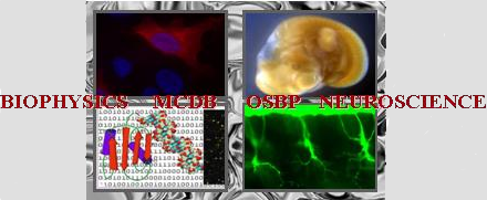
Poster abstracts
Poster number 88 submitted by Nicklaus Halloy
Core clock timing properties in the PS19 mouse model of tauopathy
Nicklaus R. Halloy (Neuroscience Graduate Program, The Ohio State University), Megan Formanowicz (Division of Pharmaceutics and Pharmacology, The Ohio State University), Nguyen Nhi Lien Pham (Department of Neuroscience, The Ohio State University), Kari R. Hoyt, Karl Obrietan (Department of Neuroscience, The Ohio State University)
Abstract:
Circadian disruptions are frequently reported in neurodegenerative disorders, including Alzheimers disease and frontotemporal dementia; the accumulation of microtubule-associated protein tau (MAPT) into oligomers and neurofibrillary tangles, a pathological hallmark of these conditions, may underlie those abnormalities. The link between pathological tau aggregation and circadian dysfunction is not well understood, especially regarding the role of the suprachiasmatic nucleus (SCN), the principal circadian pacemaker. To examine whether pathological tau within the SCN alters its structure or timing, we performed a histological and functional characterization of the PS19 (Prnp-huMAPT*P301S) transgenic mouse model of tauopathy. Histologically, hyperphosphorylated tau was evident within the SCN of PS19 mice early in disease (aged 2 months) and localized to both major SCN neuronal subpopulations: vasopressin-expressing neurons in the SCN shell and vasoactive intestinal peptide-expressing neurons in the SCN core. Functionally, we profiled daily locomotor activity as a measure of SCN clock timing and phasing in male PS19 (n = 8) and wild-type (WT; n = 8) mice from 3 to 11 months of age and in female PS19 (n = 6) and WT (n = 6) mice from 7 to 11 months of age. Analyses of overall activity patterns, rates of re-entrainment to shifted light/dark cycles, and intrinsic circadian timing revealed no significant differences between PS19 and WT animals, indicating preserved clock timing and entrainment properties of the SCN despite tau pathology. To probe SCN timing at the molecular level, we generated a PS19::Per1-Venus fluorescent clock reporter line and applied a tau-fibril seeding protocol to induce tau pathology in SCN tissue. Despite evidence of robust seeding-induced tau aggregation, in vitro fluorescence-imagingbased profiling detected no effect on clock periodicity. Together, these findings indicate that in the PS19 tauopathy model, tau aggregation within the SCN is not sufficient to disrupt SCN molecular timing properties. These findings raise the possibility that circadian disturbances observed in tauopathy patients arise from dysregulation of
Keywords: circadian, tauopathy, SCN